colocalize.com© 2008
Reasons for using RBNCC
RBNCC produces the correct answer.
No other method does - unless the images are totally free from noise.
Research is costly, why blow the investment with incorrect image analysis.
RBNCC eliminates the contribution of both signal-dependant Poisson noise and signal-independant background noise.
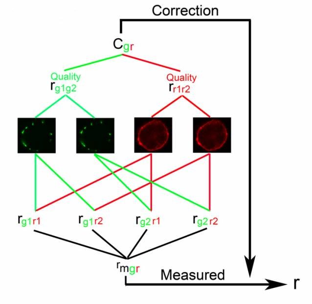
RBNCC is easy to implement. All it requires is four images and six measurements.
RBNCC makes efficient use of photons, enabling fast, repeated imaging and the effective use of quickly bleaching fluorophores.
Image quality can be estimated from the correlation coefficients between the individual fluorophores.
RBNCC produces the correct answer. Unbeatable!
Outline of RBNCC
Acquire a pair of images for each fluorophore.
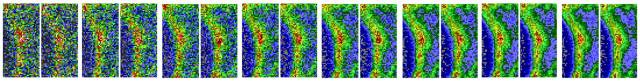
Make a noise estimate for each fluorophore by measuring the autocorrelation between the image pairs. In an ideal world two images of the same stationary object should correlate perfectly, a correlation of 1.000. An autocorrelation of less than one is due to noise and shows that the image quality is not perfect.
The quality estimates for the two fluorophores are combined into an overall correction factor.
Measure the mean correlation between the four possible between fluorophore correlations.
Apply the correction factor to the measured correlation between the two fluorophores to generate a correlation that is independant of noise.
References where RBNCC was used to measure colocalization:
Li, D., Ropert, N., Koulakoff, A., Giaume, C. & Oheim, M. (2008) Lysosomes are the major vesicular compartment undergoing Ca2+-regulated exocytosis from cortical astrocytes. J. Neurosci. 28, 7648-7658
Luccardini, C. et al. (2009) Measuring mitochondrial and cytoplasmic Ca2+ in EGFP expressing cells with a low-affinity calcium Ruby and its dextran conjugate. Cell Calcium 45, 275-283
Mahammad, S. et al. (2010) Limited cholesterol depletion causes aggregation of plasma membrane lipid rafts inducing T cell activation. Biochim. Biophys. Acta. 1801, 625-634. Article.
Waskova-Arnostova, P. et al. (2013) Right-to-left ventricular differences in the expression of mitochondrial hexokinase and phosphorylation of Akt. Cell Physiol. Biochem. 31, 66-79
Dinic, J. et al. (2015) The T cell receptor resides in ordered plasma membrane domains that aggregate upon patching of the receptor. Sci. Rep. 5:10082. Article.
Boulding et al. (2016) Differential roles for DUSP family members in epithelial-to-mesenchymal transition and cancer stem cell regulation in breast cancer. PLoS One 11:e0148065